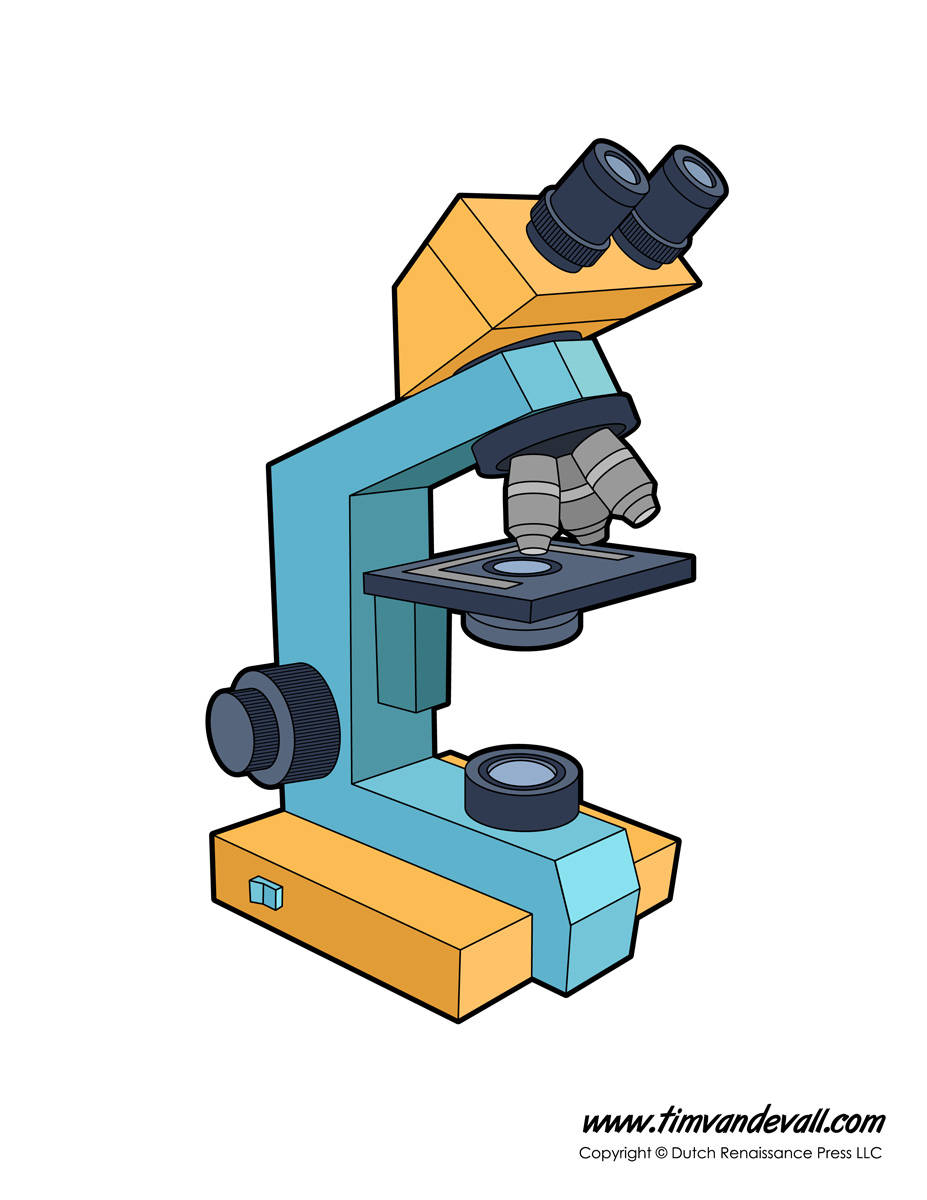
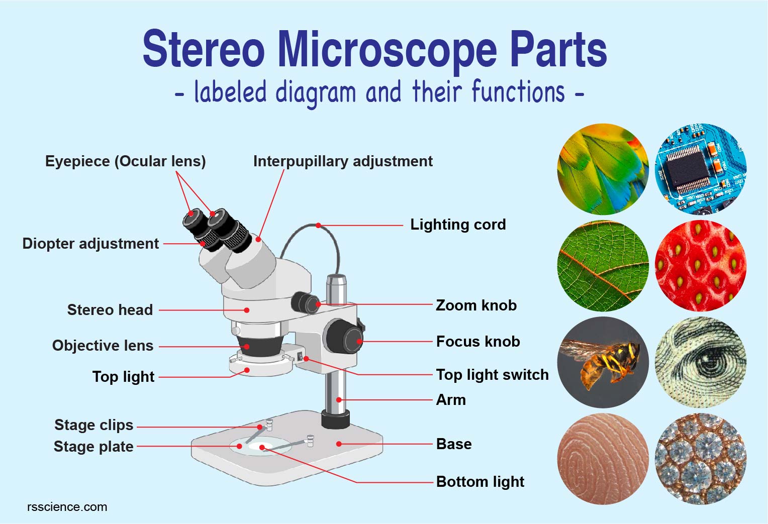
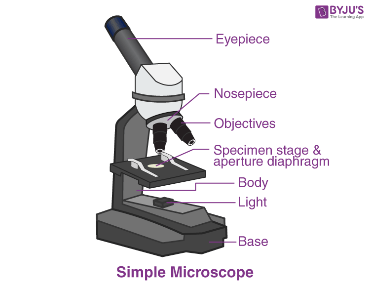
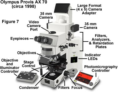
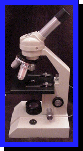
43 light microscope with labels
PDF Label parts of the Microscope Label parts of the Microscope: . Created Date: 20150715115425Z Compound Light Microscopes | Products | Leica Microsystems Leica Microsystems provides microscope parts and accessories to perfectly tailor your imaging system for your needs and budget. Darkfield Microscopes A darkfield microscope offers a way to view the structures of many types of biological specimens in greater contrast without the need of stains. Products Leica Science Lab Key Questions Meet Mica
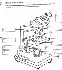
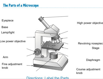
Microscope Labeling - The Biology Corner This simple worksheet pairs with a lesson on the light microscope, where beginning biology students learn the parts of the light microscope and the steps needed to focus a slide under high power. The labeling worksheet could be used as a quiz or as part of direct instruction where students label the microscope as you go over what each part is used for.
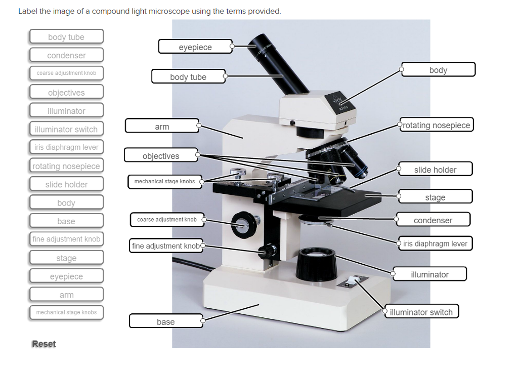
Light microscope with labels
Parts of the Microscope with Labeling (also Free Printouts) Home Microscopes Parts of the Microscope with Labeling (also Free Printouts) By Editorial Team March 7, 2022 A microscope is one of the invaluable tools in the laboratory setting. It is used to observe things that cannot be seen by the naked eye. Table of Contents 1. Eyepiece 2. Body tube/Head 3. Turret/Nose piece 4. Objective lenses 5. › microscopy › enZEISS Axio Observer for Life Science Research Fast switchable light sources and filters give you highest spectral flexibility and speed. Select the ideal camera to always get the image quality and speed your applications require. Whether keeping your sample in focus for long-term imaging or adapting your objective to your sample, it's all automatic with this highly organized system. Student's Guide: How to Use a Light Microscope An ultraviolet microscope uses UV light to view specimens at a resolution that isn't possible with the common brightfield microscope. It utilizes UV optics, light sources, as well as cameras. Because of the shorter wavelengths of UV light (180-400 nm), the image produced is clearer and more distinct at a magnification approximately double what ...
Light microscope with labels. Compound Light Microscope Optics, Magnification and Uses In order to ascertain the total magnification when viewing an image with a compound light microscope, take the power of the objective lens which is at 4x, 10x or 40x and multiply it by the power of the eyepiece which is typically 10x. Therefore, a 10x eyepiece used with a 40X objective lens, will produce a magnification of 400X. › microscopy › enZEISS Lightsheet 7 – Light Sheet Microscope Which optical clearing method you choose will depend on the tissue, your fluorescent labels, and the size of the sample. Lightsheet 7 is designed to match all these conditions. Image specimens at up to 2 cm in size at any refractive index between 1.33 and 1.58, and in almost all clearing solutions. Light Microscope- Definition, Principle, Types, Parts, Labeled Diagram ... Brightfield Light Microscope (Compound light microscope) This is the most basic optical Microscope used in microbiology laboratories which produces a dark image against a bright background. Made up of two lenses, it is widely used to view plant and animal cell organelles including some parasites such as Paramecium after staining with basic stains. Label the microscope — Science Learning Hub All microscopes share features in common. In this interactive, you can label the different parts ...
Light Microscope : Main Parts of Light Microscope | Biology Lens system in light microscope: In light microscope, the lens system is the most important. The lens systems parts are as follows — 1. Condenser: It collects and focusses light on the material. 2. Objective lens: It produces the image and also magnifies it. 3. Eyepiece or ocular lens: ADVERTISEMENTS: Compound Light Microscope: Everything You Need to Know A compound light microscope is a type of light microscope that uses a compound lens system, meaning, it operates through two sets of lenses to magnify the image of a specimen. It's an upright microscope that produces a two-dimensional image and has a higher magnification than a stereoscopic microscope. It also goes by a couple of other names ... Microscope With Labels clip art | Microscope parts, Scientific method ... Description Use this blank handout as a way for students to record microscope drawings. Aside from the drawig itself, studnts are prompted to title the drawing, include the magnification of the microscope, and give a quick description of what they are viewing. Keywords: Microscope Drawings Lab Biology. S. Learning with Shedd Aquarium. Microscope Labeling Game - PurposeGames.com This is an online quiz called Microscope Labeling Game. There is a printable worksheet available for download here so you can take the quiz with pen and paper. This quiz has tags. Click on the tags below to find other quizzes on the same subject.
› AmScope-120X-1200X-BeginnerAmazon.com: AmScope 120X-1200X 52-pcs Kids Beginner ... Nov 18, 2013 · Buy AmScope 120X-1200X 52-pcs Kids Beginner Microscope STEM Kit with Metal Body Microscope, Plastic Slides, LED Light and Carrying Box (M30-ABS-KT51),Black: Microscopes - Amazon.com FREE DELIVERY possible on eligible purchases Labeling the Parts of the Microscope | Microscope World Resources Labeling the Parts of the Microscope This activity has been designed for use in homes and schools. Each microscope layout (both blank and the version with answers) are available as PDF downloads. You can view a more in-depth review of each part of the microscope here. Download the Label the Parts of the Microscope PDF printable version here. Compound Microscope Parts - Labeled Diagram and their Functions The illuminator provides a source of light. The light is focused by the condenser and passing through the specimen placed on the stage. The light is then collected and formed into an image by an objective lens. We see the magnified images through the eyepiece. A clear image needs perfect focusing by adjusting the coarse and fine focus knobs. Light microscopes - Cell structure - Edexcel - BBC Bitesize Microscopes are used to produce magnified images. There are two main types of microscope: light microscopes are used to study living cells and for regular use when relatively low magnification and...
› microscopy › enZEISS LSM 900 with Airyscan 2 - Compact Confocal Microscope ... Airyscan 2 is an area detector with 32 circularly arranged detection elements. Each of these acts as a small pinhole, contributing to super-resolution information, while the complete detector area collects more light than the standard confocal setting. This produces much greater light efficiency while capturing enhanced structural information.
Labelled Diagram Of A Light Microscope - GlobalSpec Products/Services for Labelled Diagram Of A Light Microscope Microscopes - (706 companies) ...and transmission electron microscopes. Acoustic and ultrasonic microscopes use sound waves to create images of the sample. Compound microscopes use a single light path. These types of microscopes can have a single eyepiece (monocular) or a dual eyepiece...
en.wikipedia.org › wiki › Two-photon_excitationTwo-photon excitation microscopy - Wikipedia In scattering tissue, on the other hand, the superior optical sectioning and light detection capabilities of the two-photon microscope result in better performance. Applications Main. Two-photon microscopy has been involved with numerous fields including: physiology, neurobiology, embryology and tissue engineering.
How does a Light Microscope Work [ Complete Guide ] A light microscope uses a thinly sliced specimen placed on a slide. The slide is placed on the stage. To adjust the specimen, we use a Coarse Adjustment knob. The coarse adjustment knob adjusts the stage to bring the specimen in focus. It adjusts the stage by either moving the stage up and down or moving the body tube.
› NATIONAL-GEOGRAPHIC-Dual-StudentNational Geographic Dual LED Student Microscope Aug 07, 2017 · Buy NATIONAL GEOGRAPHIC Dual LED Student Microscope - 50+ pc Science Kit with 10 Prepared Biological & 10 Blank Slides, Lab Shrimp Experiment, Perfect for School Laboratory, Homeschool & Home Education: Microscopes - Amazon.com FREE DELIVERY possible on eligible purchases
Simple Microscope - Diagram (Parts labelled), Principle, Formula and Uses The magnification power of a simple microscope is expressed as: M = 1 + D/F Where M = Magnification power D = the lease possible distance of distinct vision of eye, typically 25cm F = Focal length of the convex lens It is to be noted that
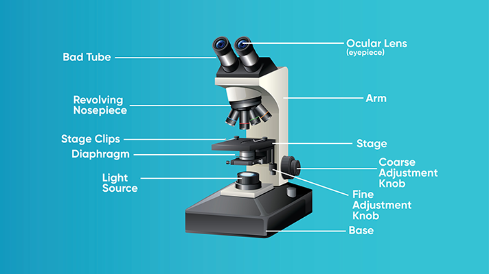
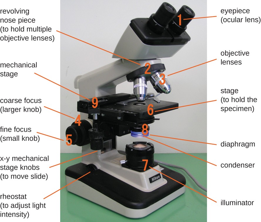
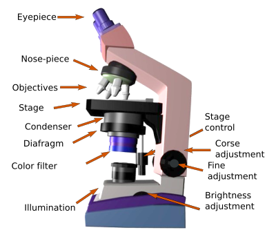
Microscope, Microscope Parts, Labeled Diagram, and Functions Revolving Nosepiece or Turret: Turret is the part of the microscope that holds two or multiple objective lenses and helps to rotate objective lenses and also helps to easily change power. Objective Lenses: Three are 3 or 4 objective lenses on a microscope. The objective lenses almost always consist of 4x, 10x, 40x and 100x powers. The most common eyepiece lens is 10x and when it coupled with ...
Parts of a microscope with functions and labeled diagram - Microbe Notes The higher the magnification of the condenser, the more the image clarity. More sophisticated microscopes come with an Abbe condenser that has a high magnification of about 1000X. Diaphragm - it's also known as the iris. Its found under the stage of the microscope and its primary role is to control the amount of light that reaches the specimen.
www2.nau.edu › lrm22 › lessonsMicroscopy - Northern Arizona University Microscope Drawings. When drawing what you see under the microscope, follow the format shown below. It is important to include a figure label and a subject title above the image. The species name (and common name if there is one) and the magnification at which you were viewing the object should be written below the image.
Compound Microscope Parts, Functions, and Labeled Diagram Compound Microscope Definitions for Labels. Eyepiece (ocular lens) with or without Pointer: The part that is looked through at the top of the compound microscope. Eyepieces typically have a magnification between 5x & 30x. Monocular or Binocular Head: Structural support that holds & connects the eyepieces to the objective lenses.
A Study of the Microscope and its Functions With a Labeled Diagram Light Microscopes: These use light rays to illuminate objects. e.g. Dissection microscopes and compound microscopes. Electron Microscopes: These illuminate objects with a beam of highly charged electrons. e.g. Transmission electron microscope (TEM) and scanning electron microscope (SEM).
Label the light microscope | Teaching Resources Label the light microscope Subject: Biology Age range: 11-14 Resource type: Worksheet/Activity 0 reviews File previews pdf, 96.38 KB Low ability worksheet, label the main parts of a microscope, key terms given at the bottom of the worksheet. Creative Commons "Attribution" Report this resource to let us know if it violates our terms and conditions.
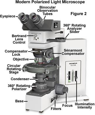
Polarized Light Microscopy - Microscope Configuration | Olympus LS Condensers for Polarized Light Microscopy. Basic substage condenser construction in a polarized light microscope is no different from an ordinary condenser used in brightfield microscopy. In all forms of microscopy, the degree of condenser optical correction should be consistent with that of the objectives. Typical laboratory polarizing ...
Light Microscope Parts, Function & Uses - Study.com Anton van Leeuwenhoek (1632-1723) invented a simple (one-lens) microscope around 1670. Leeuwenhoek made lenses by carefully grinding and polishing solid glass to make his microscopes.
Light Microscope: Functions, Parts and How to Use It To use a light microscope, you can follow the steps below carefully. Start with a low lens and a clean slide. The microscope stage should be lowered as low as possible. Center the slide so that the specimen is under the objective lens. Use the coarse adjustment knob to get a general focus. Then slowly move up the stage until focus is achieved.
Microscope Parts and Functions The specimen is placed on the glass and a cover slip is placed over the specimen. This allows the slide to be easily inserted or removed from the microscope. It also allows the specimen to be labeled, transported, and stored without damage. Stage: The flat platform where the slide is placed.
Label the Light Microscope - Labelled diagram - Wordwall Drag and drop the pins to their correct place on the image.. Eyepiece, Light Source, Base, Stage, Stage Clips, Fine Focus, Coarse Focus, Arm, Objective Lens.
An Introduction to the Light Microscope, Light Microscopy Techniques ... Fluorescence microscopy is used to image samples that fluoresce, that is, they emit long-wavelength light when illuminated with light of a shorter wavelength. Examples include biological samples that are intrinsically fluorescent or have been labeled with a fluorescent marker, as well as single molecules and other nanoscale fluorophores.
Student's Guide: How to Use a Light Microscope An ultraviolet microscope uses UV light to view specimens at a resolution that isn't possible with the common brightfield microscope. It utilizes UV optics, light sources, as well as cameras. Because of the shorter wavelengths of UV light (180-400 nm), the image produced is clearer and more distinct at a magnification approximately double what ...
› microscopy › enZEISS Axio Observer for Life Science Research Fast switchable light sources and filters give you highest spectral flexibility and speed. Select the ideal camera to always get the image quality and speed your applications require. Whether keeping your sample in focus for long-term imaging or adapting your objective to your sample, it's all automatic with this highly organized system.
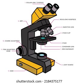
Parts of the Microscope with Labeling (also Free Printouts) Home Microscopes Parts of the Microscope with Labeling (also Free Printouts) By Editorial Team March 7, 2022 A microscope is one of the invaluable tools in the laboratory setting. It is used to observe things that cannot be seen by the naked eye. Table of Contents 1. Eyepiece 2. Body tube/Head 3. Turret/Nose piece 4. Objective lenses 5.
















Post a Comment for "43 light microscope with labels"